Images of Melanoma in People with Skin of Color

What Does Melanoma Look Like on a Variety of Skin Tones?
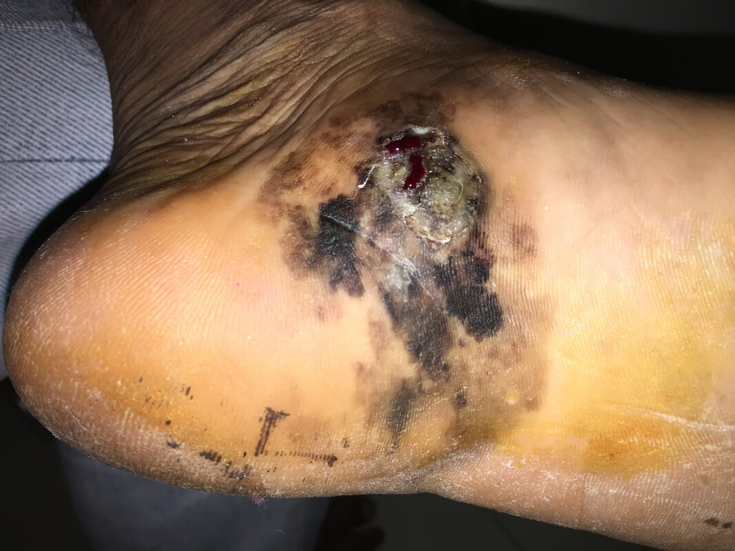
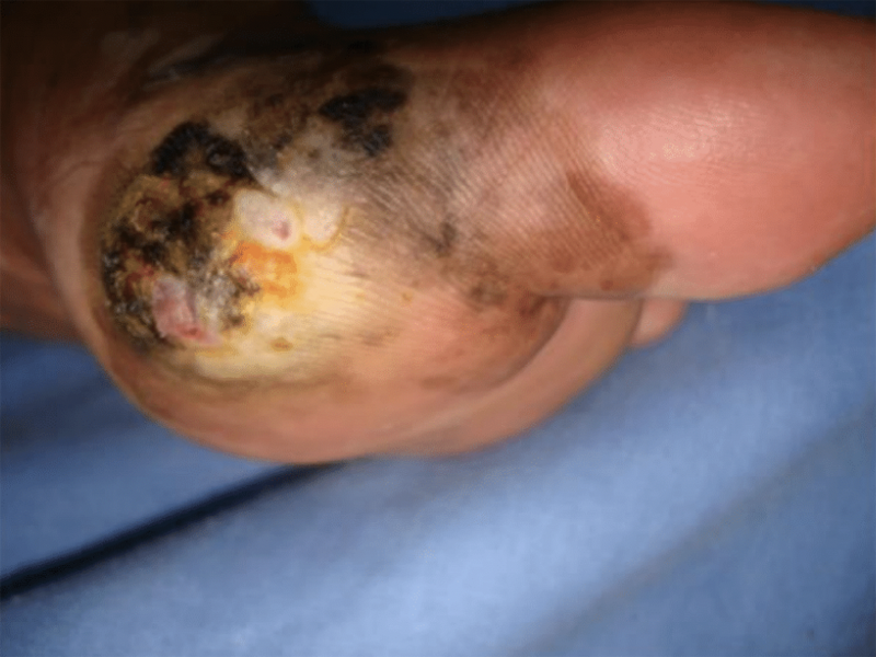
Melanoma is a potentially life-threatening cancer affecting people of all skin tones. The way it presents itself in individuals with dark skin can be different from those with medium or lighter complexions. For example, individuals with medium complexions may notice an unusual-looking mole whereas darker complexions may notice a wound.
Some people with skin of color may have a misconception they are not at risk for developing melanoma due to their natural protection from UV rays. Although it is true that melanin pigment offers some protection, it is not usually much, and it does not prevent cancer. A misconception about this fact can lead to delayed diagnosis and treatment.
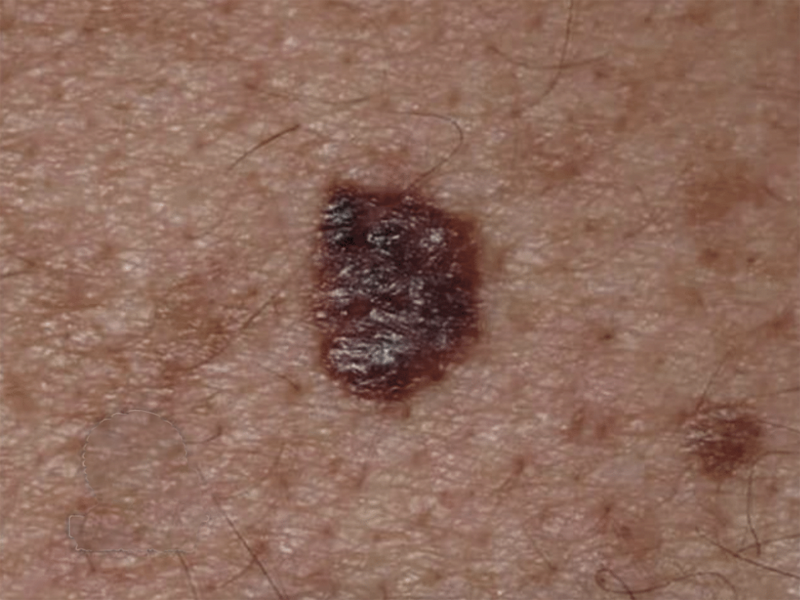
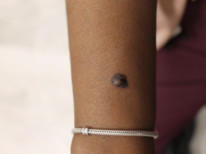
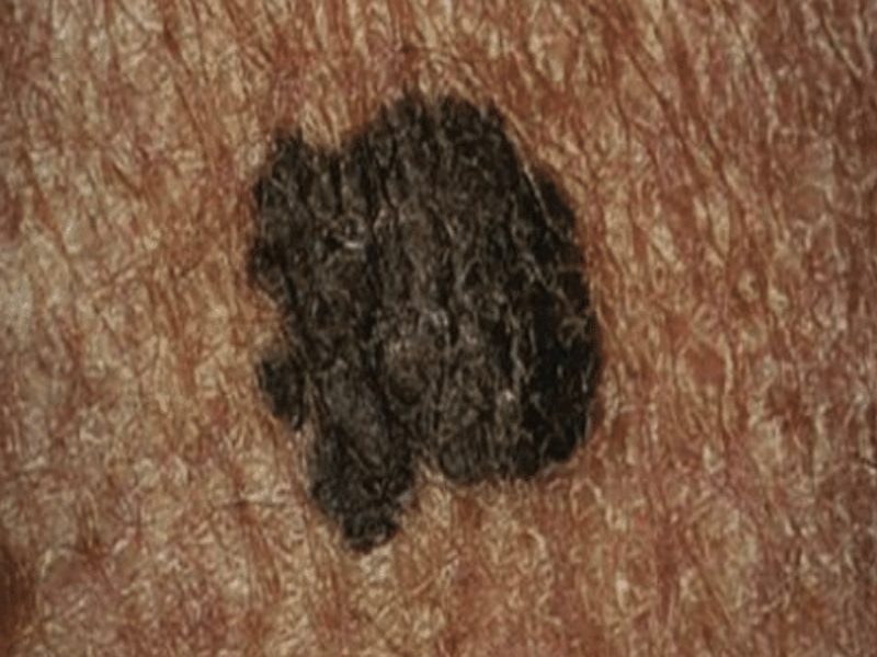
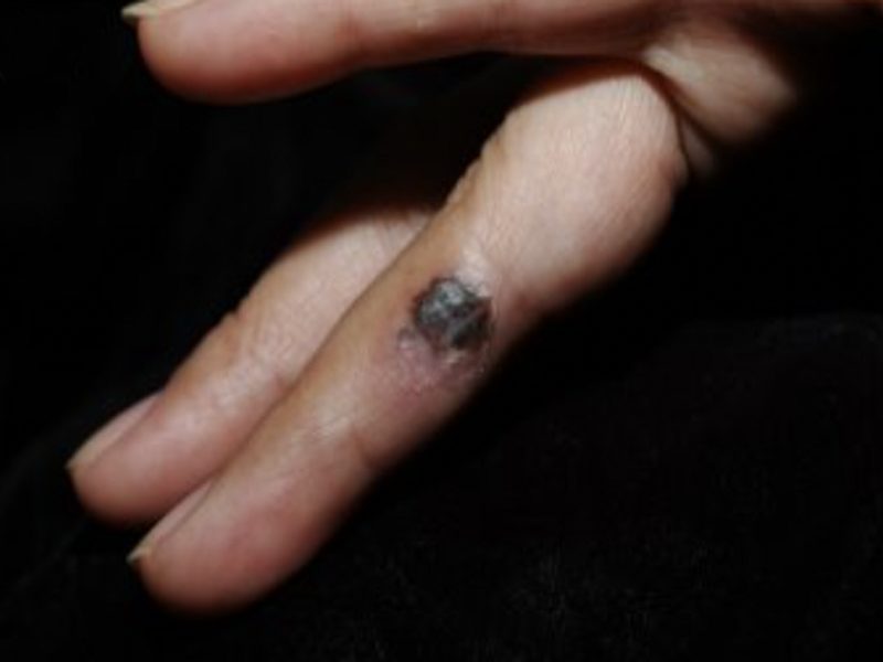
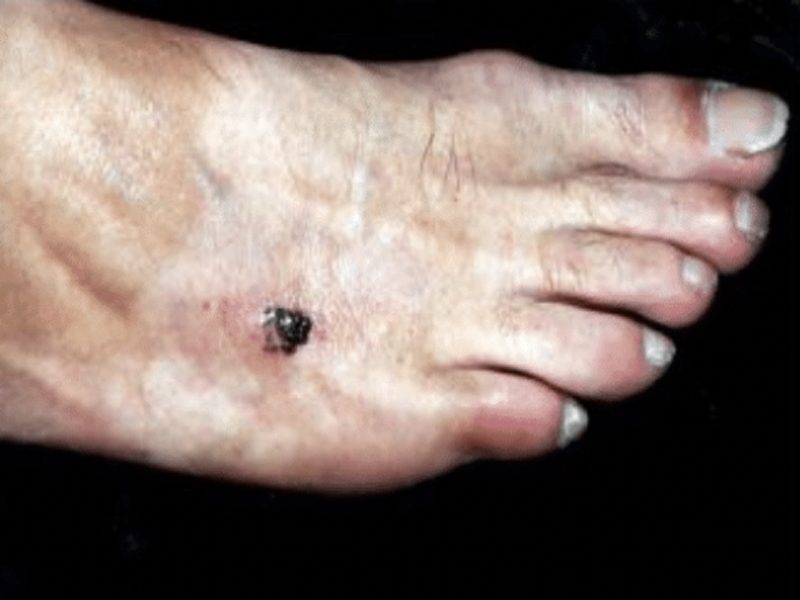
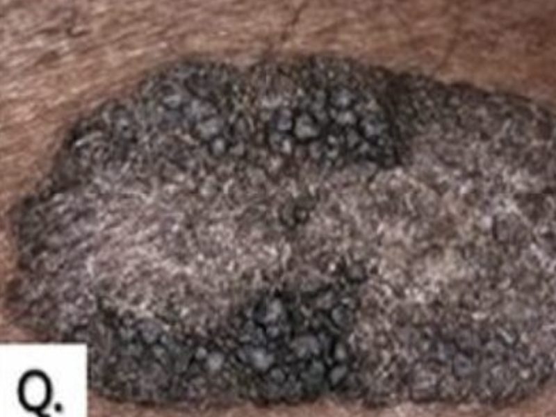
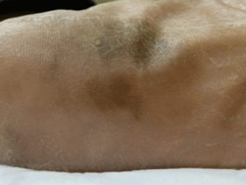
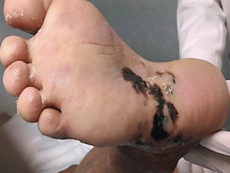
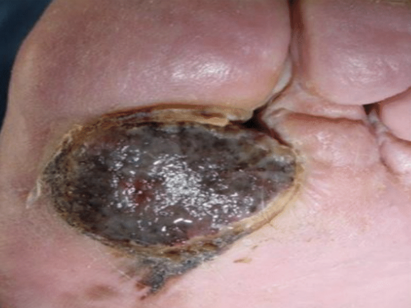
Melanoma in people with medium or darker skin tones may appear as dark brown or black spots, which can be mistaken for a benign growth. Additionally, melanoma may develop on areas of the body that are not typically exposed to the sun, such as the palms of the hands, soles of the feet or inside the mouth.
It is important for all people to educate themselves on what melanoma looks like on someone with their skin tone. Routine self-examinations and screenings by a dermatologist aid early detection and intervention, and it’s especially important that your dermatologist has experience identifying skin cancer in people with skin of color. Being proactive reduces the risk of developing advanced melanoma.
In general, when looking at spots to identify potential cancer, it’s helpful to ask yourself:
- Is the lesion new?
- Has the lesion changed or grown over time?
- Does the area appear unusual compared to your other skin?
- Does the lesion bleed for a while or never seem to heal?
- Has someone suggested you get it checked?
These are all signs that you should have your skin checked by a healthcare provider. And don’t wait: The earlier melanoma is diagnosed, the easier it is to treat.
How People of all Skin Tones Can Develop Melanoma
To help raise awareness about the importance of early detection and diagnosis of melanoma, we’ve compiled images of lesions in a range of individuals, ethnicities, and complexions. These images are meant to serve as a visual guide for recognizing potentially concerning spots on your skin. However, it is important to note that no image bank can be comprehensive, and you may have a suspicious spot that does not resemble any of the images provided.
If you notice any spot on your skin that concerns you, whether it looks like the images provided or not, it is crucial to make an appointment with a dermatologist for further evaluation.
Skin of color is broadly defined as racial and ethnic groups with skin tones darker than individuals who inherited a majority of Northern European genetic ancestry. The scientific and medical communities in the U.S. usually include some or all of the following groups when defining skin of color: American Indian/Alaska Native, Asian/Pacific Islander, Black, Hispanic/Latino/Latina and other non-White individuals that have mixed ethnicities and races.
Melanoma Photo Gallery
Melanoma looks different between individuals with variations in skin tone. More importantly, it also develops in different areas of the body. The photos appearing in this gallery are designed to provide a range of melanoma representations on different skin tones from many groups in the U.S., regardless of the amount of melanin or pigment in the cells. It also highlights the different areas of presentation. Please note that your skin and/or a suspicious lesion may look different from the images here. If you have a question about any lesion on your skin, see your dermatologist or healthcare provider.
While melanoma looks different on different people and different skin tones, here are some things to look for on your skin:
- New or existing moles or lesions that are asymmetrical, have an irregular border, are more than one color, are larger than a pencil eraser, or have changed in any way
- A dark, rapidly spreading patch on the skin
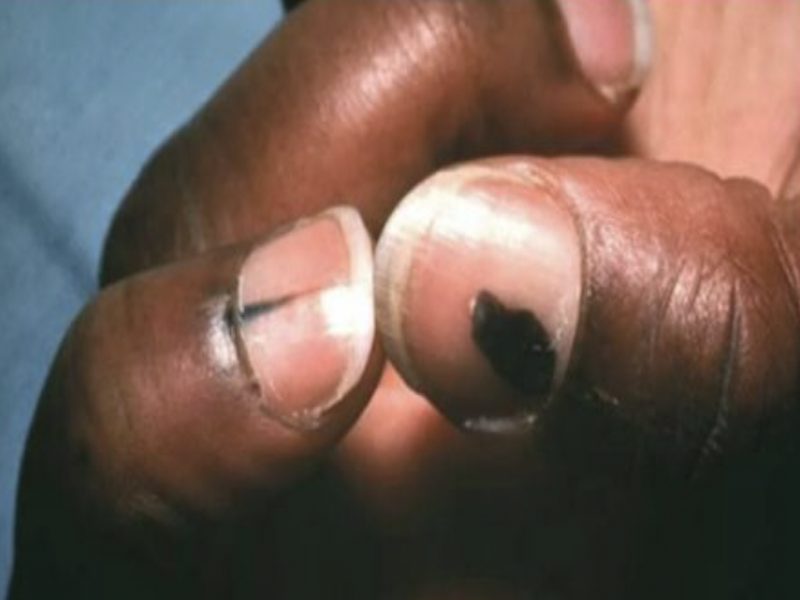
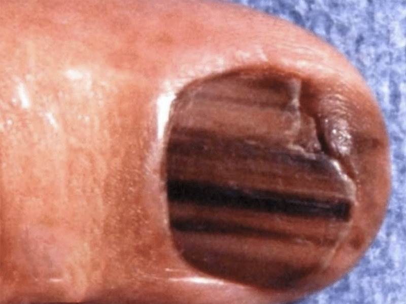
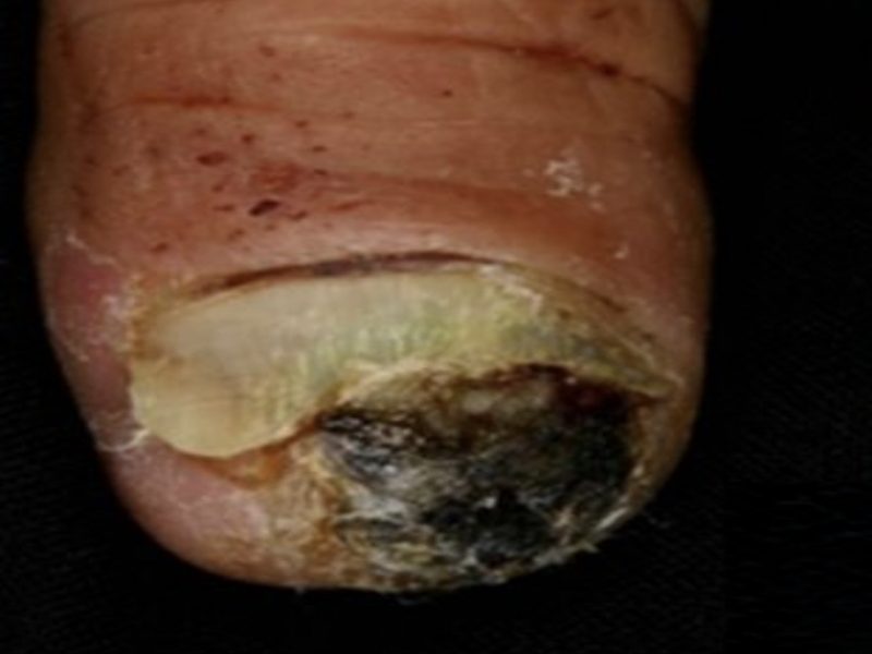
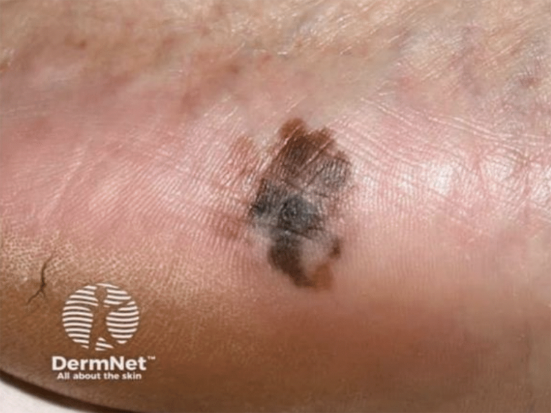
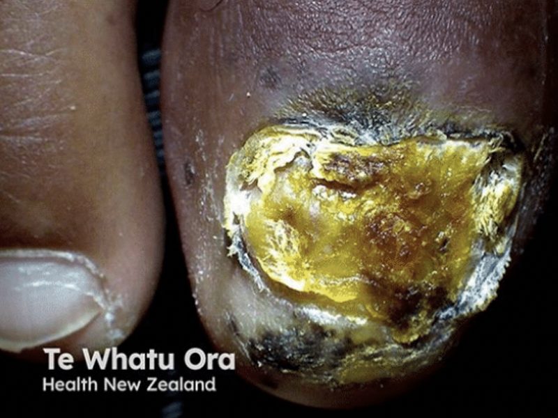
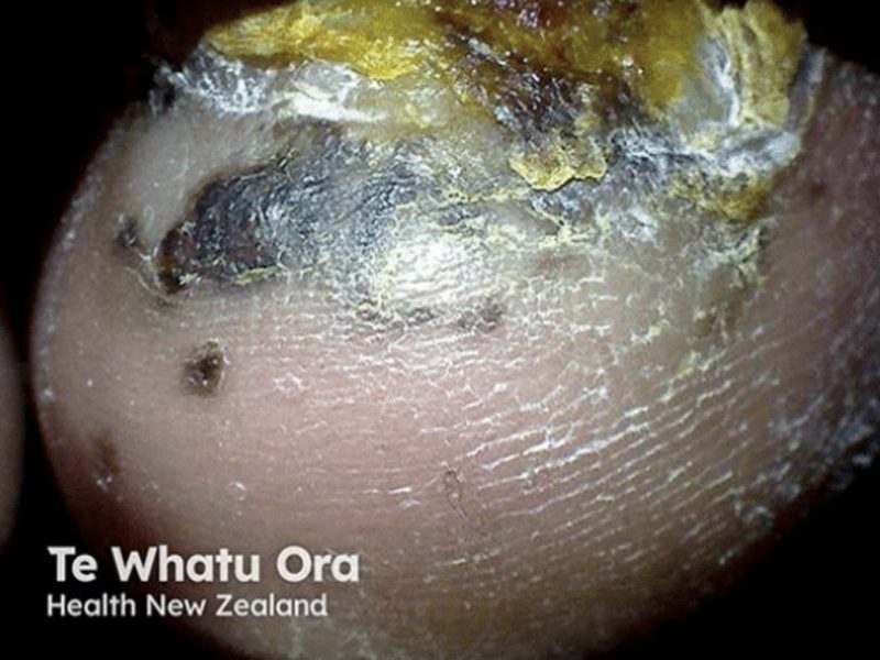
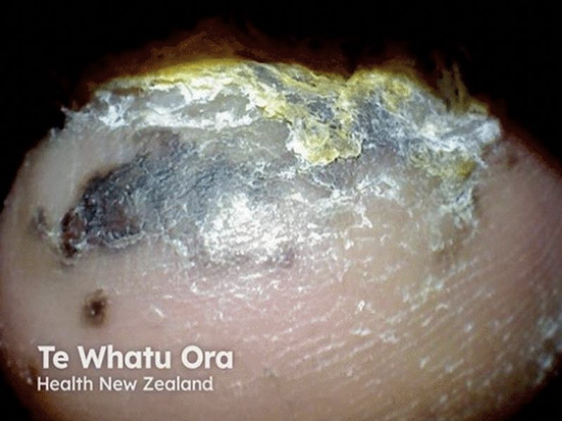
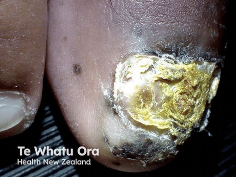
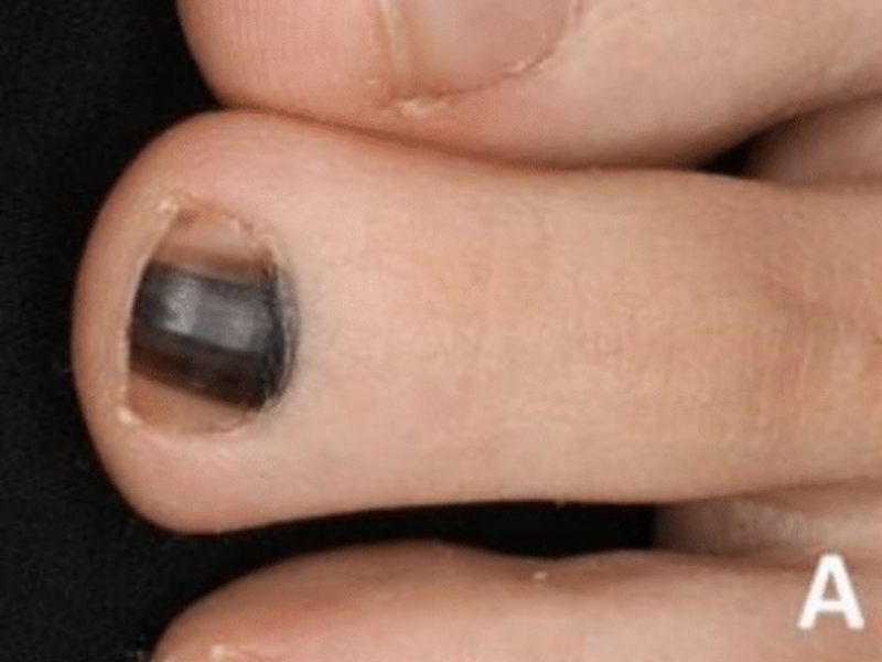
- An irregularly shaped growth with unusual color or an elevated, thickened patch on the palms of hands or soles of the feet; a streak under a nail
- A dark-colored lesion in the mouth, nose, or genital areas
Source: Dr. Steve Daveluy , MD, FAAD
Source: Dr. Carl Washington, MD, and Dr. Mona Saraiya, MD, MPH. The Centers for Disease Control and Prevention
Source: Dr. Carl Washington, MD, and Dr. Mona Saraiya, MD, MPH. The Centers for Disease Control and Prevention
Source: Kim SH & Tsao H. Biomolecules . 2025;15(1):120. doi : 10.3390/biom15010120
Source: Kim SH & Tsao H. Biomolecules . 2025;15(1):120. doi:10.3390/biom15010120

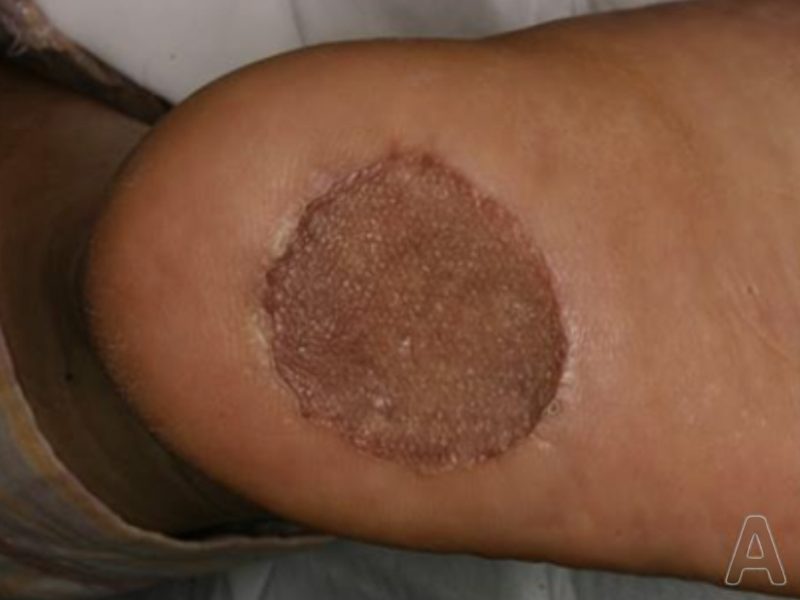
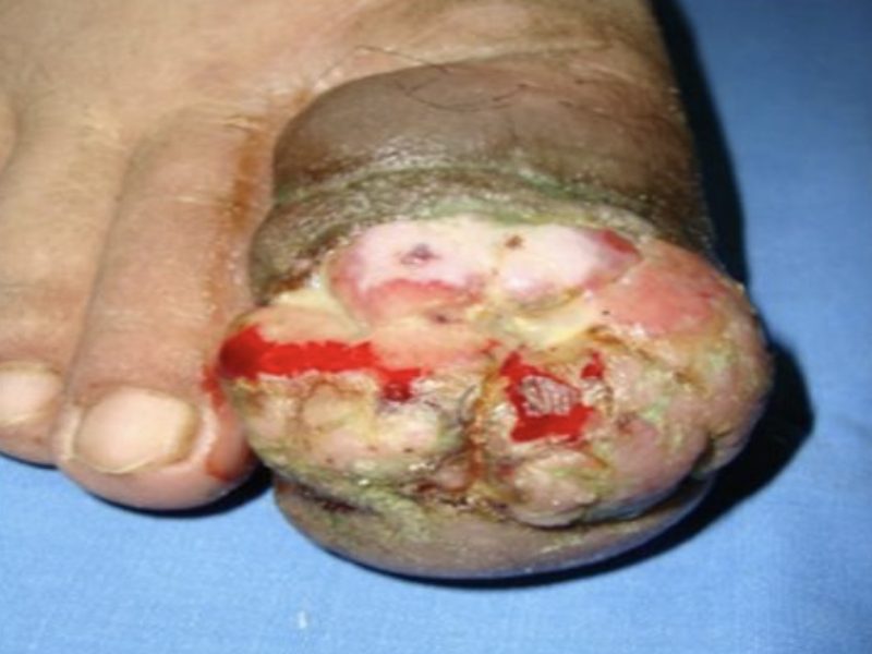
lentiginous melanoma on the palm
The purple line was drawn by the surgeon before the procedure, wide local excision, was used to remove the lesion.
Source:
Nakamura Y, Teramoto Y, Sato S, et al. Current Surgical Management of Acral Lentiginous Melanoma.2015. Intech. doi: 10.5772/59133
Source: DermNet New Zealand
Source:
Nakamura Y, Teramoto Y, Sato S, et al. Current SurgicalManagement of Acral Lentiginous Melanoma. 2015. Intech.
doi:10.5772/59133
Source: Diehl K, Stoos E, Becker A et al.
Front Med (Lausanne).2024;11:1500216. doi: 10.3389/fmed.2024.1500216.
eCollection 2024
Source: Nakamura Y, Teramoto Y, Sato S, et al. Current Surgical Management of
Acral Lentiginous Melanoma. 2015.
Intech.doi: 10.5772/59133
Source: DermNet New Zealand
Source:https://doi.org/10.1126/sciadv.abq6147
Source:
https://doi.org/10.1126/sciadv.abq6147
Source: Barra, Martínez R, Herrera- González NE,Fernández Ramírez F, et al. Acral Melanoma — A Distinct Molecular and Clinical Subtype. 2015.
Intech. doi : 10.5772/59093
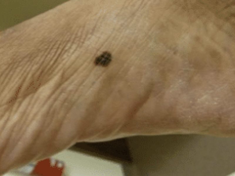
likely melanoma
Source: Shutterstock
Source:
Barra-Martínez R, Herrera-
González NE,Fernández
Ramírez F, et al. Acral Melanoma—A DistinctMolecular and Clinical Subtype. 2015. Intech.doi: 10.5772/59093
Source: Barra-Martínez R, Herrera-
González NE, Fernández-
Ramírez F, et al. Acral Melanoma—
A Distinct Molecular and
Clinical Subtype. 2015.
Intech.doi : 10.5772/59093
Source: DermNet New Zealand
Source: DermNet New Zealand
Source: DermNet New Zealand
Source: DermNet New Zealand
Source: Nakamura Y, Teramoto Y, Sato S, et al. Current Surgical Management of Acral Lentiginous Melanoma. 2015.
Intech. doi : 10.5772/59133
Source: Barra- Martínez R, Herrera- González NE, Fernández- Ramírez F, et al. Acral Melanoma — A Distinct Molecular and Clinical Subtype. 2015.
Intech. doi : 10.5772/59093