How Ocular Melanoma is Diagnosed

Can eye doctors see ocular melanoma?
Eye melanomas can be detected during a routine dilated eye exam in people before any symptoms have developed. After dilating, or temporarily widening, the pupil using eye drops, the eye doctor can examine eye structures using a standard lighted instrument called an ophthalmoscope. They may also use an indirect ophthalmoscope or slit lamp or a specialized instrument called a gonioscopy lens to detect tumors growing in parts of the eye that are difficult to see. Ocular melanoma can also be detected in specialized pictures of the eye that are sometimes taken during routine eye exams to assess general eye health. The same testing is done if a patient presents with new ocular symptoms or has a freckle that has been followed over time.
Although a suspected melanoma can be detected during a routine eye exam, additional specific tests are usually done to confirm a diagnosis and rule out other conditions.

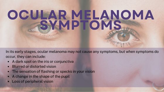
See an ophthalmologist immediately if you have any of the symptoms listed above.
What tests are done to diagnose ocular melanoma?
Following detection of a suspected melanoma during a routine eye exam, people with suspected ocular melanoma may be referred to a type of doctor called an ocular oncologist who specializes in eye cancer to confirm the diagnosis and discuss a treatment plan. These specialist doctors diagnose ocular melanoma based on symptoms, patient history, clinical evaluation, and specialized test results.
A variety of tests are available to help in the diagnosis of ocular melanoma, and doctors consider various factors in selecting which tests to use in a given patient. Specifically for uveal melanoma, eye imaging tests that doctors commonly recommended to diagnose ocular melanoma include:
- Fundus photography. Special cameras are used to photograph the eye lesion to view the size, borders, and location relative to eye structures.
- Ocular ultrasound. A wand-like instrument held against the eye sends sound waves through the eye to create a picture of the eye and tumor mass.
- Optical coherence tomography. Similar to an ocular ultrasound, but this test uses light instead of sound waves to create pictures of the back of the eye, including the uveal tract and retina.
- Angiography. To view the blood vessels in the back of the eye, including those around the tumor, a colored or fluorescent dye is injected into a vein (usually in the arm). The dye travels to the blood vessels of the eye and a special camera is used that can detect and photograph the dye to create pictures of the eye. This test can be used to check if the symptoms are caused by other common eye conditions instead of melanoma.
Doctors diagnose many types of cancer by taking a biopsy (removing a small piece of tumor for laboratory testing). However, a biopsy is not usually needed to diagnose uveal melanoma. A diagnosis of uveal melanoma can be confirmed by the eye exam and imaging tests above. But a biopsy is sometimes helpful to check for certain gene changes that predict spread (read more about how doctors can predict uveal melanoma spread here). Depending on the tumor location, the tumor sample can be collected in different ways, including a fine needle aspiration or biopsy, where a very thin needle is used to remove a small sample of the tumor. Sometimes a biopsy is done after treatment (for example, if the tumor is removed by surgery). Liquid biopsies, where tumor cells are collected from a blood sample instead of directly from the tumor, are becoming more common but are currently mainly done as part of clinical trials. This type of biopsy may be useful to check for metastatic spread without needing to make a cut or insert a needle into the eye.
After an ocular melanoma is diagnosed, additional tests may be recommended to check to see if the melanoma has spread to other parts of the body.
Conjunctival melanomas are managed much more like skin melanoma. The primary management is surgical removal. The tumor being removed is then sent to a pathologist to make sure the surgeon was able to fully remove the tumor.
How is metastatic ocular melanoma diagnosed?
Standard imaging tests are used to detect ocular melanoma that has spread to other parts of the body. These tests can include:
- Ultrasound of the abdomen to look for melanoma growth in the liver.
- Chest x-ray to check for melanoma in the lungs.
- Computer tomography (CT) scan to detect cancer growing into nearby structures around the eye or spread to other organs, for example, the liver
- Magnetic resonance imaging (MRI) to look for tumor growth outside of the eye, for example in the liver.
Blood tests may be done after someone is diagnosed with eye melanoma. For people with uveal melanoma, blood tests commonly include liver function tests because problems with liver function can be a sign that the cancer has spread to the liver.
What are the most common symptoms that the disease has spread? What are the main symptoms of metastatic uveal melanoma?
The symptoms of metastatic uveal melanoma depend on which part(s) of the body the cancer has spread to and the degree of spreading.
What do stages mean regarding the spread of metastatic ocular melanoma?
Ocular melanoma is classified into prognostic stages based on the size of the original tumor, how much it has grown into nearby areas, and whether it has spread to other parts of the body, including the lymph nodes or distant organs.
Doctors classify uveal and conjunctival melanoma into stages that are associated with better or worse prognosis based on specific criteria set by a professional organization called the American Joint Committee on Cancer (AJCC) called the TNM system. The stages are based on the size of the eye tumor and how much it has grown into surrounding tissues (the “T” in TNM), whether the melanoma is growing in nearby lymph nodes (“N”), and whether the melanoma has spread to other parts of the body, or metastasized (“M”). Stage I disease has the best prognosis, while Stage IV disease is associated with the worst prognosis. Some stages are further divided into sub-categories (A, B, C) representing progressively worse prognosis within a stage.
The TNM criteria that doctors use to stage ocular melanoma depend on whether the melanoma is uveal or conjunctival, and, for uveal melanoma, where within the uveal tract it is located. In general, Stage I, II, or III ocular melanomas have not spread widely outside of the eye, while Stage IV melanomas are growing in distant parts of the body.
In general, for the most common ocular melanomas (ciliary body and choroid uveal melanomas),
- Stage I: the smallest-sized tumors with no growth into the ciliary body or outside of the eye
- Stage II: either (Stage IIA) smallest-sized tumors growing into the ciliary body or with a small outgrowth reaching outside of the eye (smaller than 5 mm) OR small-to-medium-sized (Stage IIB) or medium-sized (Stage IIC) tumors with no growth into the ciliary body or outside of the eye
- Stage III: small-to-medium-sized (Stage IIIA), medium-sized (Stage IIIB), or large (Stage IIIC) tumors growing into the ciliary body or with a small outgrowth reaching outside of the eye (smaller than 5 mm) OR large tumors with no growth into the ciliary body or outside of the eye (Stage IIIB) OR any tumors with larger growths extending outside the eye (more than 5 mm; Stage IIIC)
- Stage IV: the melanoma has spread outside of the eye and is growing in areas of the orbit that do not touch the eye, in the lymph nodes, and/or in distant organs like the liver or lungs
What is the most likely location for metastasis from ocular melanoma?
Uveal melanoma usually spreads either by growing directly into nearby tissues or by entering the blood vessels to reach distant parts of the body. It is relatively uncommon for this type of melanoma to spread to the lymph nodes.
The most common location for uveal melanoma to spread to is the liver. The liver is affected in up to about 90% of metastatic cases. Other organs including the lungs, skin, soft tissues, and bone, may also be affected. However, the liver is usually the first part of the body to be affected in people whose melanoma has spread to more than one organ.
Conjunctival melanoma can also enter the bloodstream and spread to distant parts of the body, most commonly to the lung, brain, liver, skin, bone, or gastrointestinal tract. Unlike uveal melanoma, this type of ocular melanoma also commonly spreads to the regional lymph nodes.
Read more about factors that predict ocular melanoma spread here.

